The nucleus of the phagocytic cell is not visible in this section. (The ones that look like squashed spirals.) Just below this group of degenerating axons is part of a phagocytic cell (it could be a macrophage or a Schwann cell) engulfing several bundles of myelin debris. I can see a close grouping of 4 large prominent degenerating myelinated axons in the upper right quadrant of the photograph. To me, they have a very obvious crumpled appearance.

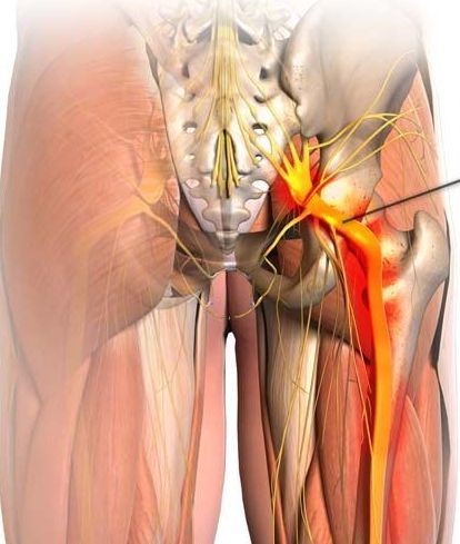
There are lots of degenerating axons in this photograph. The sciatic nerve may be injured high along its course at the roots and plexus level underneath the pyriformis muscle in the buttock (most notoriously by injury from intramuscular injections) and along its entire course in the thigh by fracture, missile wounds orother types of injuries.This is actually a pretty good image, although it would be easier to interpret in color since the lipid in the myelin looks black while proteins in the cytoplasm are more of a dark blue. Sciatic Entrapment, Compression Or Injury Sites The stimulating electrode must be a needle electrode over the sciatic notch, which is halfway between the ischial tuberosity and greater trochanter. Place the recording electrodes on those muscles used in peroneal or posterior tibial testing. The posterior tibial nerve may be involved as part of a sciatic nerve injury at the popliteal fossa in the tarsal tunnel following ankle injury and rarely at an anterior opening of the abductor hallucis muscle. Femoral Entrapment, Compression Or Injury Sites


You can also stimulate it above the inguinal ligament.īecause this nerve is difficult to stimulate in obese patients, especially above the inguinal ligament, needle electrodes may be used for that purpose.

Stimulate the nerve in the groin over the femoral triangle or at Hunter’s canal. Place the active recording electrode over the vastus medialis muscle and the reference electrode on the patella.


 0 kommentar(er)
